Apr 2015 : Report of KCIAPM Slide Seminar
R V Metropolis, Bengaluru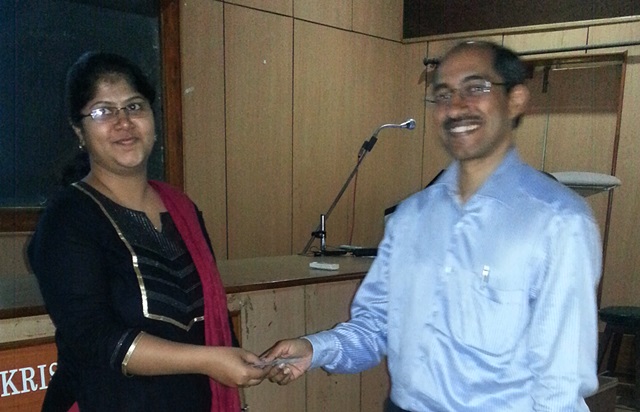
The second slide seminar of 2015 was hosted by RV Metropolis Lab. The moderators were- Dr Ravikumar H N , Dr VaniRavikumar and Dr Santosh K.V.
Ten histopathology cases were selected from the archives of RV Metropolis and forty sets of slides were despatched to various medical colleges of Karnataka. Ten PGs from different medical colleges were selected to present the microscopic features of these cases. Also, microphotographs of these cases were posted in KCIAPM web site.
Eighteen colleges sent in their responses, which were compiled by the moderators.
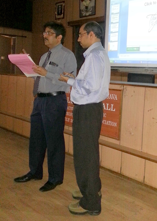
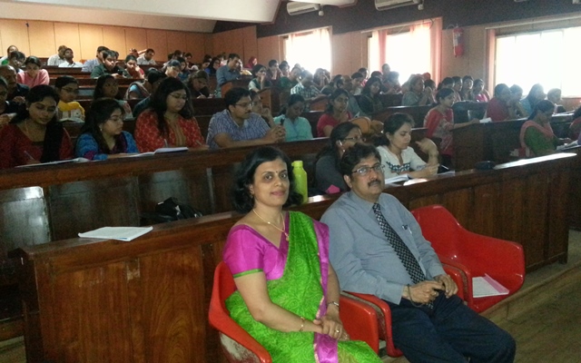
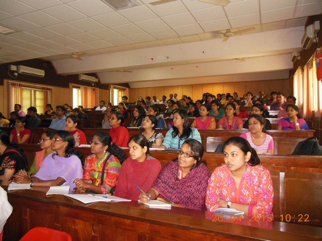
Breakfast was hosted by RV Metropolis Lab and the session started at 10 AM. Dr Vijay shankara s , Hon Secretary began the session by welcoming the students.Case 1-3 discussed by Dr Ravikumar, Cases 4-7 by Dr VaniRavikumar and cases 8-10 by Dr Santosh KV. A total of 120 delegates attended this program. The venue was Pathology Lecture Hall at Victoria Hospital, BMC&RI. Dr Raghupathi, Prof&HOD, was gracious to provide the venue.
The discussion emphasised on approach to case diagnosis, with relevant recent classifications, role of IHC markers and special stains. Basics in approach to a histopathlogic diagnosis were discussed in detail. There was active participation by the PGs and it was truly an interactive slide seminar.
The session ended at 1.30PM. The best presenter was Dr Anoosha from Adichunchanagiri Institute of Medical sciences, B G Nagara and she was awarded a cash prize by the moderators.
Dr Anoosha receiving the “Best Presenter’ Award from Dr Santhosh.
The feedback from the PGs was that it was a very useful seminar and helped them for a practical approach to slide interpretation. The consultants from RV Metropolis have reiterated that they will host such slide seminars every year and keep up the tradition as well their commitment to teaching PG students.
Case Slide Diagnosis
| Histopath No | Presenters | History | Diagnosis |
|---|---|---|---|
| H72374 | Dr. Aneesha Asok Kumar MSRMC | 58/F. Mass in the right nasal cavity. Fragmented bits received. C/S - homogenous grey-white | Plasmablastic NHL |
| H72126 | Dr. Shashikala MIMS MANDYA | 65/M with generalised lymphadenopathy and splenomegaly. Section from axillary lymph node. | Castleman’s disease progressing to low progressing to low grade NHL, with sarcoid-like granulomas |
| H75688 | Dr. Ankit Malhotra KIMS, | 31/F. Left superficial parotidectomy specimen. C/S - 2.3x2 cm nodule. | Pleomorphic RMS adenoma with stromal atypia |
| H47605 | Dr. Nitika Grover VIMS, Ballari | 50/F. Wide excision specimen of left thigh tumor. Mass measured 15x14x5 cm; had grey-white fleshy cut surface | Pleomorphic RMS |
| H74295 | Dr. Keerthi RKMIO, | 45/F. Left radical nephrectomy specimen, 21x12x10 cm. C/S grey-white with hemorrhage and necrosis. | Chromophobe RCC |
| H76289 | Dr. Anoosha K AIMS, BG NAGARA | 31/M. Left suprarenal mass. Gross specimen - 7x5x3.5 cm, cut surface - yellowish | Adrenal Oncocytoma |
| H76324 | Dr. Najmunnissa S MVJMC | 21/M. History of left nasal obstruction since childhood. Multiple fragments of bony & soft tissue sent. | Psammomatoid ossifying fibroma |
| H77464 | Dr. Ajaz M Dar Yenepoya MC | 75/M. Excision biopsy from swelling in right lateral thigh. C/S - grey-white nodule 4.5x4 cm | Giant solitary trichoepithelioma |
| H70248 | Dr. Narasimha Murthy BMCRI | 53/F. Right ovarian cystectomy. Specimen was 17x13x10 cm. C/S - solid and cystic areas. | Adult granulosa cell tumor |
| H74326 | Dr. Prahi Kukreja KS Hegde MC | 58/F with external hemorrhoids. Hemorrhoidectomy specimen. | Pemphigus vulgaris with hemorrhoids |